
By Dr. Luke Haydock (University of Guelph)
On the 7th of January, 2022, Dr. Charles Schwarten kicked off a new year of Davis-Thompson Foundation Friday seminars – and what better way to start the new year than sinking our teeth into a crash course on equine dental histology?! The topics covered in this webinar were of great interest to me and to many others in the audience, so I would like to extend my immense gratitude to Dr. Schwarten and colleagues for advancing our understanding in this often-perplexing area of pathology.
As promised, Dr. Schwarten began his talk by providing us with a truly foundational summary and review of “normal” equine dental development, anatomy, and histology. 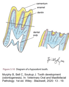 Complete with extremely detailed figures, I found my understanding of the base-truths of equine dental pathology to be much advanced by Dr. Schwarten’s explanations of odontogenesis, normal equine dentition, and normal gross anatomy and microanatomy. This was a highly detailed discussion that simply cannot be missed, and I strongly encourage you to check out the talk as it was delivered straight from the horse’s mouth, so to speak!. A complete recording of the webinar can be found for 6 months on the Davis-Thompson Foundation YouTube page (link: https://www.youtube.com/watch?v=M-kM1r8pmMU).
I personally would like to commend Dr. Schwarten on providing such a thorough and foundational review of “normal”, because it really provided much more context for when the discussion moved on to the “abnormal”. As teeth are so dissimilar to most other tissues in the body, those uninitiated to the world of dental pathology will really benefit from Dr. Schwarten’s ground-up approach to the recognition of dental lesions. After reviewing the normal microanatomy of teeth, discussion turned to some of the features seen in teeth as they adapt to physiological wear-and-tear (e.g. secondary dentin), as they respond to injury / disease (e.g. tertiary dentin), and which are seen incidentally (e.g. pulp stones).
With this solid foundation established, Dr. Schwarten, in collaboration with his colleagues at the Joint Pathology Center (JPC), was then kind enough to share some recent findings in the evaluation of equine dental disease. Based on submissions to JPC, the most common entities diagnosed in equine dental submission are, in descending order, equine odontoclastic tooth resorption and hypercementosis (EOTRH) (37.2 %), periodontitis (30.7 %), pulpitis / pulp necrosis (29.5 %), reactive hypercementosis (20.5 %), caries (17.9 %), fracture (12. 8 %), bacterial infection (12. 8 %), pulp stones (5.1 %), odontodysplasia (2.6 %), ameloblastoma (1.3 %), and squamous cell carcinoma (1.3 %). A brief review of some of these common entities followed (i.e. EOTRH, caries, tooth root infections), particular focus was dedicated to EOTRH – briefly, EOTRH is a progressive condition of incisors and canine teeth that is most commonly diagnosed in middle-aged to older horses. 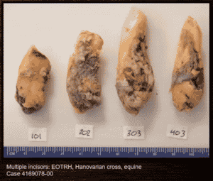 It is a painful condition typified by grossly appreciable swelling upon clinical examination that tends to begin on the outer incisors and progress mesially. Histologically, it is characterized by lysis of both dentin and cementum with concurrent excessive deposition of cementum – concurrent periodontal and / or endodontal disease may also be present. The pathogenesis of this disease is unclear, but it has been hypothesised that it may be related to the increased occlusive angle of incisors with increasing age. I found the discussion of these entities to be of great interest, and I once again encourage the reader to check them out for themselves on the foundation YouTube page.
And as if we weren’t already spoiled, the Q&A session that wrapped up this session was rich with yet more expert insights and hot tips – Dr. Schwarten provided a thorough yet concise review of his methods for preparing teeth for histological evaluation. The histological preparation of teeth can be a daunting task at the best of times, so this discussion was of much interest to me and many others in the audience. These methods were also reviewed in a poster presentation by Dr. Schwarten and colleagues at last year’s American College of Veterinary Pathologists Annual Meeting (Cudd et al., 2021 – Histologic Findings in Equine Teeth Submitted from and Equine Referral Hospital) – check it out too!
Once again, on behalf of the Davis-Thompson Foundation, I would like to extend my immense gratitude to Dr. Schwarten for volunteering to deliver this webinar. I thoroughly enjoyed this talk, as I’m sure all those in attendance did. The global veterinary pathology community continues to benefit enormously from passionate and diligent professionals such as him who are willing to contribute to the effort in advancing education in veterinary pathology around the world.